Copyright © 2007-2018 Russ Dewey

The Brain
Your brain is a true wonder of nature, probably the only one that wonders about itself. Humans take the brain for granted because it is part of their basic package, but brains are amazing things. The human brain is the most complex piece of matter in the known universe.
Sitting behind the protective shell of the skull, cushioned in a special liquid called cerebrospinal fluid, the brain is a living system composed of many billions of individual nerve cells called neurons. Each neuron is a highly complex system in itself, surrounded by even larger numbers of glial cells that play a supporting role.
Estimates of the total number of neurons in the human brain vary greatly. The number, from 20 billion to a trillion, depends on which cells are included.
Glial cells number about 60 billion in the cerebral cortex. They are now thought to modify information processing, not just metabolic functions of neurons, so they should be counted, too.
What structures make up the central nervous system?
The human nervous system consists of two main parts: (1) the central nervous system (CNS) consisting of the brain and spinal cord, and (2) the peripheral nervous system (PNS), consisting of nerves outside the brain and spinal cord, traveling to every part of the body.
The brain is pink and soft while living. Brain surgeons say it has the consistency of thick custard. Only when preserved for observation does the brain appear gray and claylike.
Very conspicuous on the brain are large wrinkles or convolutions. These convolutions vastly increase the surface area of the brain.
CNS Development in the Embryo
The human nervous system starts as a tube of cells in the developing embryo. Over the first few weeks of development, the front part of the tube thickens. Already the three basic divisions of the central nervous system are visible: the forebrain, midbrain, and hindbrain.
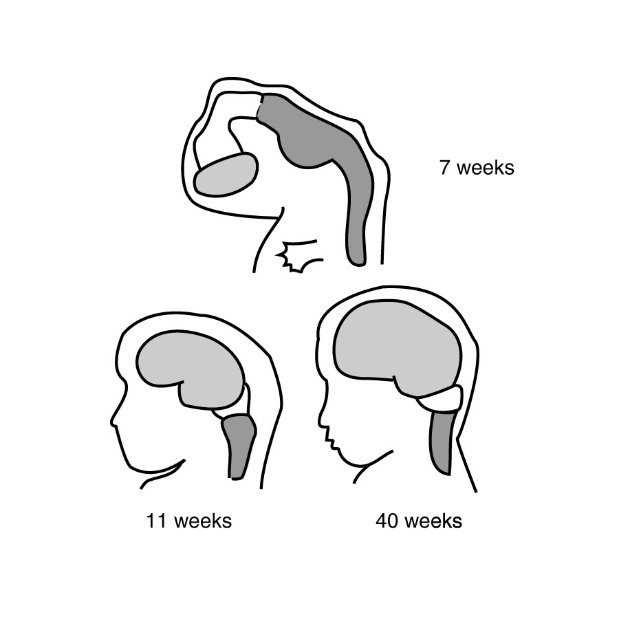
CNS Development in the Embryo
Late in embryological development the convolutions begin to form, giving the human brain its distinctive wrinkled appearance. The convolutions continue to form and deepen for years after a baby is born.
The convolutions form because the rapidly growing brain has different layers of cells that grow at different rates, causing buckling. Because of this folding, the brain appears to have distinct parts called lobes.
Fissures–deep folds in the surface of the brain–mark off each lobe. The opposite of a fissure is a gyrus (plural gyri ). A gyrus is an outward fold similar to a hilltop on the brain. The following figure shows major gyri on the human brain.
What causes convolutions to form? What is a fissure? A gyrus?
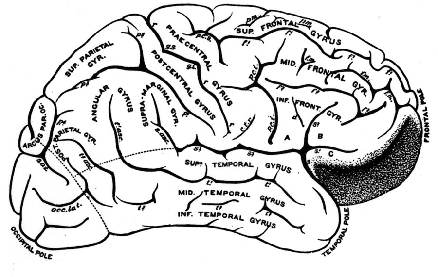
Gyri on the brain–prefrontal areas in black
The cerebrum (pronounced either SARAH-brum or suh-REE-brum) is the topmost part of the brain. It consists of everything you see in this figure, which is facing right. The prefrontal area, darkened above for emphasis, is an important area for higher-level thought processes in humans.
The Cerebral Cortex
The cerebrum's outermost layer is called the cerebral cortex. This layer averages 3.2 mm thick in human beings, less than a quarter inch. The layer of cortex covers the surface of the cerebrum and goes down into the fissures.
The word cortex means rind or shell in Latin. It refers to a dense outer layer of neurons on any brain structure, not just the cerebrum. (Smaller brain structures like the thalamus and cerebellum also have cortical layers.)
What is the cerebral cortex? What does "cortex" mean?
When a brain is preserved, cortical areas–areas of cortex–appear gray because they contain dense concentrations of cell bodies. This is called gray matter ("grey" matter in most English-speaking countries; "gray" is the American spelling).
Technically, gray matter refers only to cortical areas. Other brain tissue is called white matter. It consists of communication lines: neural fibers called axons which run from the cells in the cortical layers to other parts of the brain. The axons appear white in these areas because they are coated with a fatty substance, myelin.
What makes gray matter gray and white matter white?
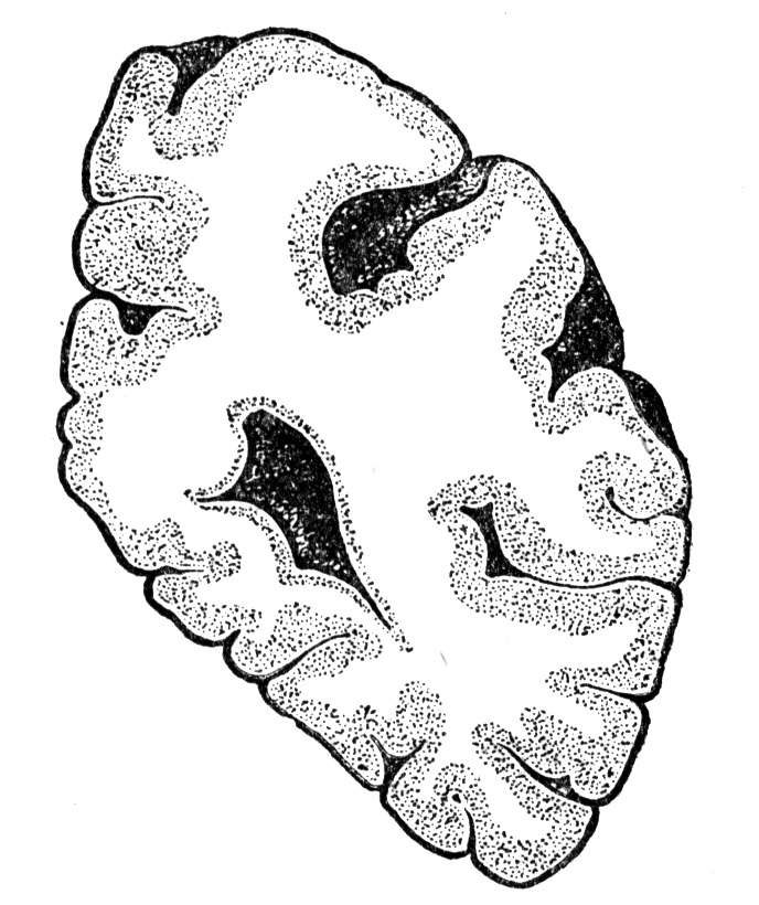
Gray and White Matter
A slice of brain tissue is shown above. You can see how the gray matter extends down into the fissures. This is significant because it creates a large surface area for nerve cell bodies. The cerebral cortex, if unfolded, would have about the same surface area as a small umbrella.
How large is the cortical area, if unfolded?
The cerebral cortex has many specialized areas that work together in ordinary thought processes. Brain scans typically show several widely separated areas of the cortex active simultaneously when a mental task is performed.
The cerebral cortex is important to humans, but people can live without major portions of it, especially if the damage is done early in life. Babies are sometimes born with half the cerebral cortex missing, yet they grow up with normal mental abilities.
Hydrocephalics can suffer compression of brain tissue. "One of these patients gained a first-class honors degree (Sheffield University) in mathematics and is socially completely normal. And yet...has virtually no brain" (Lewin, 1980, p.1232, quoted in Maratsos and Matheny, 1994).
Lobes of the Brain
Lobes are large areas of the cerebral cortex bounded by fissures in the brain. At the back of the brain is the occipital [ok-SIP-it-al] lobe. Normally the occipital lobe is specialized for vision.
In blind people the neural tissue in the occipital lobes may be used for other functions such as touch and hearing (Amedi, Merabet, Bermpohl, and Pascual-Leone, 2005). That illustrates the plasticity or adaptability of brain tissue.
Correlations between brain areas and functions can change. Unused areas commonly become involved in new functions.
At the back of the occipital lobe is the primary visual cortex, the first stop for visual information entering the cortex. Stimulating the primary visual cortex with a weak electric current causes phosphenes: little glowing lights in the visual field.
What visual event is caused by stimulation of the back of the occipital lobe?
Once there was hope that implanting an array of electrodes in the occipital lobe would allow messages to be flashed to blind people like words on an electronic billboard. However, it turned out stimulation at a particular location did not always cause a phosphene in the same place, so those hopes were abandoned.
More complex visual areas extend beyond the primary visual cortex into the temporal lobe on the side of the brain. Stimulating this area can result in visual hallucinations. Damage along the lower side of the temporal lobe can cause visual agnosia–inability to recognize objects.
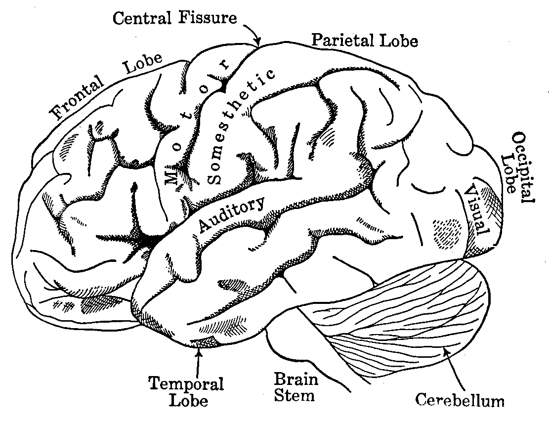
Major lobes on a brain "facing left" (frontal lobes to left)
The parietal (par-EYE-it all) lobe is the middle area of the brain. Parietal cortex in the right hemisphere is usually devoted to spatial processing. That is the talent used to figure out maps or find your way around a complex environment.
The temporal lobe is on the side of the brain, below a major fold (the lateral or Sylvian fissure). The temporal lobe contains not only the secondary visual areas but also some tissue devoted to hearing, and it is involved in complex forms of information processing.
Stimulating the upper surface of the temporal lobe can result in the déjà vu sensation (the sensation of having seen something before). Stimulating another area produces profound feelings of what is real and true similar to religious ecstasy. (Ramachandran, Hirstein, Armel, Tecoma, and Iragui, 1998).
Where is the temporal lobe located? What are complex forms of information linked to the temporal lobe?
The frontal lobes are in the front of the brain. The frontal lobe is larger in humans than any other animal. The prefrontal areas right behind the eyes are involved in executive functions such as selective attention and task management (Smith and Jonides, 1999).
When the frontal lobes are damaged or isolated from the rest of the brain, people lose their ability to plan for the future and perform creative activities. This was discovered in the 1940s and 1950s when surgical operations called lobotomies were performed on some patients for pain relief. We will discuss lobotomies later in the chapter.
What are some functions supported by the frontal lobes? Where are the prefrontal areas?
Running from the top of the head down toward the ear is the central fissure or fissure of Rolando. The area behind the central fissure is called the sensory cortex, and the area in front is called the motor cortex.
Those labels are from the 1800s. Today's neuroscientists say both areas, so-called sensory and motor cortex, contain sensory and motor functions.
What is located on either side of the central fissure? What are "motor functions"?
Both areas are involved in receiving inputs from the senses (sensory functions). Both are involved in sending outputs to the muscles (motor functions).
The Homunculus
Scientists can map which parts of the brain control various parts of the body. The mapping is done by stimulating the sensory or motor cortex with a weak electric current. The stimulation often produces tingling or movement in part of the body.
Humans put great emphasis on speech and manipulation of objects by the hands, so humans have large amounts of cortex devoted to mouth, tongue, and hands. Different species have different patterns. Rats get a lot of information from their whiskers, so they have large amounts of sensory cortex devoted to their whiskers.
The following diagram represents a slice of cortex near the fissure of Rolando, running from the top of the head down toward the ear. The diagram indicates the location and amount of cortex devoted to each part of the body.
For example, just above the lateral fissure by the ear, stimulation produces a swallowing reflex. At a location on the top of the head, stimulation results in toe movement.
The map of brain connections to the body, in this particular strip of cortex, is depicted below. The amount of cortical tissue is represented by the size of the body part in the diagram.
How is the homunculus mapped out?
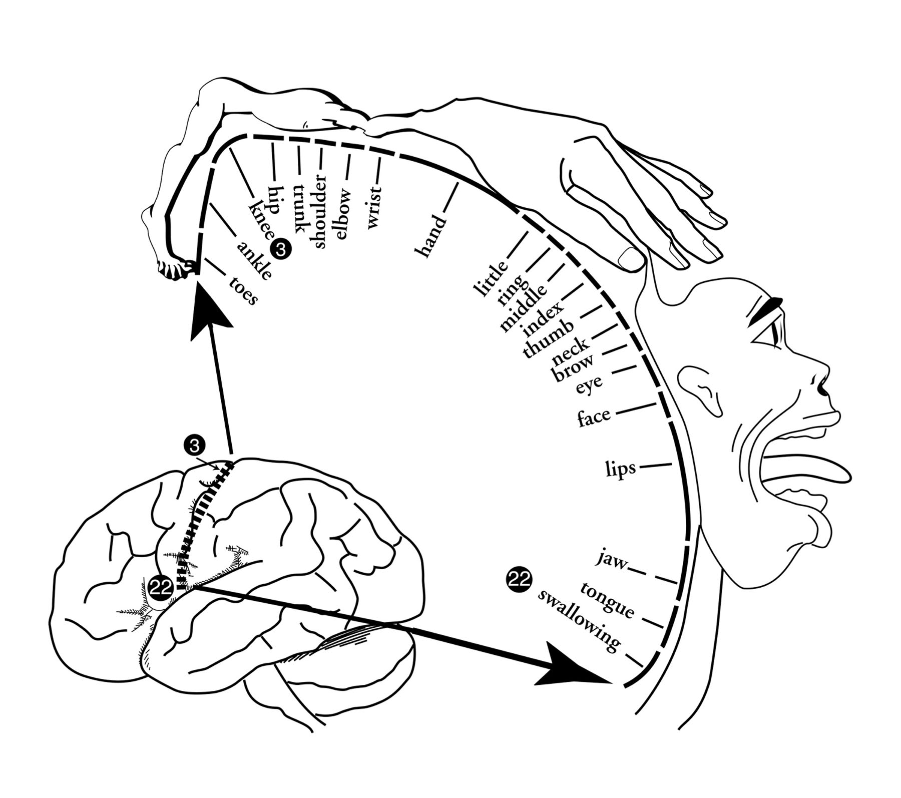
The Homunculus, based on Penfield's classic diagram
The diagram looks a bit like a grotesque little man, so it is called the homunculus (ho-MUN-q-lus) which means "little man" in Latin. The first homunculus diagram was drawn by brain surgeon Wilder Penfield in the 1940s.
Notice that the hands, lips, and tongue are large. That is because the figure is drawn to reflect how much cortex is devoted to each area of the body.
The homunculus is a textbook diagram, certainly is not a self or center of consciousness in the brain. However, humorous references to the homunculus as a little person in the head are common among psychologists.
One psychologist might say to another, "But how exactly is this mental activity carried out? Does the homunculus do it?" This is a way of saying, "You have not given us an adequate explanation!"
There are many homunculi in the brain, if the word refers to an area of cortex where body surfaces are mapped. Such maps can change with experience.
People who read Braille (which is done with an index finger) develop large areas responsive to stimulation from the index finger. A homunculus mapped on the motor cortex of such a person would have a huge index finger.
What is evidence that cortical mapping can change with experience? What therapy did this inspire?
Liepert et al. (1998) wondered if stroke patients suffering paralysis on one side might be suffering from a re-assignment of motor cortex. After a stroke, one arm might be unused for weeks or months. It might lose its representation on the motor cortex.
To encourage a revival of ability to move the paralyzed arm, Liepert and colleagues used a sling to immobilize the patient's good arm for 90% of the patient's waking hours. Each patient spent six hours a day in therapy that encouraged using the bad arm.
The result was improvement in all 73 patients treated, regardless of how much time had passed since the stroke. In fact, 30% of the patients regained full use of the formerly bad arm.
---------------------
References:
Amedi, A., Merabet, L. B., Bermpohl, F. & Pascual-Leone, A. (2005) The Occipital Cortex in the Blind: Lessons About Plasticity and Vision. Current Directions in Psychological Science, 14, 306-311.
Liepert, J., Miltner, W. H., Bauder. H., Sommer. M., Dettmers, C., Taub, E., & Weiller, C. (1998) Motor cortex plasticity during constraint-induced movement therapy in stroke patients. Neuroscience Letters, 26, 5-8.
Maratsos, M. & Matheny, L. (1994). Language specificity and elasticity: Brain and clinical syndrome studies. Annual Review of Psychology, 45, 487-516.
Ramachandran, V. S., Hirstein, W., Armel, K. C., Tecoma, E., & Iragui, V. (1998). The neural basis of religious experience. Society for Neuroscience Abstracts, 23, 519.
Smith, E. E. & Jonides, J. (1999) Storage and executive processes in the frontal lobes. Science, 283, 1657-1661.
Write to Dr. Dewey at psywww@gmail.com.
Don't see what you need? Psych Web has over 1,000 pages, so it may be elsewhere on the site. Do a site-specific Google search using the box below.